
For decades, the medical consensus surrounding the central nervous system (CNS) has been sobering: once neurons in the brain, spinal cord, or visual system are severely damaged or die off, the loss is permanent. Unlike the skin, which knits itself back together, or the liver, which can regenerate, the mammalian central nervous system has historically been viewed as a rigid, one-way street. If the delicate bridge of communication between the eye and the brain is broken, blindness or severe visual impairment follows.
However, a groundbreaking new study led by researcher Athanasios Alexandris and colleagues at Johns Hopkins University, published in JNeurosci, is actively flipping this long-held narrative on its head. Using mice as models, the research team discovered that the mammalian visual system is far more flexible, adaptable, and capable of self-healing than science previously gave it credit for.
The secret to this recovery isn't found in miraculously resurrecting dead cells, but rather in the remarkable resilience of the survivors.
The Mechanics of "Sprouting"
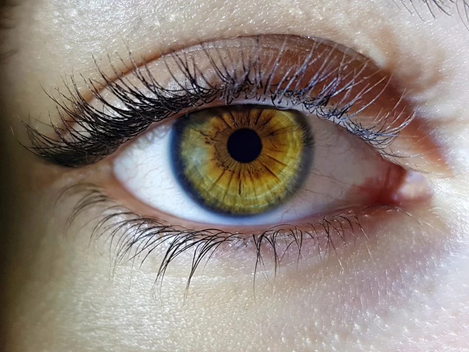
To understand the magnitude of this discovery, it is essential to look at the cellular players involved. Human and mammalian vision relies heavily on Retinal Ganglion Cells (RGCs). These are the crucial nerve cells situated near the inner surface of the retina. They act as the biological fiber-optic cables of the eye, gathering visual information and transmitting it down their long, thread-like axons through the optic nerve and into the brain for processing.
When trauma, disease (like glaucoma), or ischemic events damage these cells, many of them die. The traditional approach to reversing this vision loss focused on a seemingly impossible task: trying to coax the nervous system into growing entirely new neurons and forcing their long axons to stretch all the way back across the injury site to reconnect with their original targets in the brain.
The Johns Hopkins study upends this assumption. The researchers observed that the mammalian brain doesn't necessarily need to rebuild from scratch; instead, it looks for a detour.
The Biological Detour: When neighboring cells die, the surviving, uninjured retinal ganglion cells sense the loss. In response, they undergo a fascinating biological process called compensatory sprouting.
Think of it like a mature tree that has lost several large limbs in a violent storm. To survive and continue gathering sunlight, the tree doesn't try to instantly regrow the exact same lost limbs; instead, it sprouts dense new branches and leaves from its intact, surviving trunk and remaining branches.
Similarly, the intact neurons in the mice began growing additional branches from their existing structures—both new axons (the fibers that send signals) and new dendrites (the branches that receive signals). These new extensions physically reach out into the neural architecture to form fresh synaptic connections with other neurons. They essentially take on the workload of their fallen neighbors, creating alternative, rerouted pathways for visual signals to successfully travel from the eye to the brain.
Shifting the Paradigm of Neural Repair
This behavioral and physiological reorganization translated into highly meaningful functional improvements for the mice. While the recovery of their vision wasn't always 100% perfect, the animals regained a substantial and measurable portion of their lost visual capabilities.
This establishes concrete evidence that meaningful functional recovery is entirely possible in mammals without true neuron regrowth.
| Feature | Traditional Regeneration Model | Compensatory Sprouting Model (The New Discovery) |
|---|---|---|
| Primary Goal | Replace lost or dead neurons completely. | Maximize the utility of surviving, healthy neurons. |
| Mechanism | Forcing axons to regrow across the injury site. | Existing neurons sprout new branches to adapt. |
| Connectivity | Attempting to reconnect with original targets. | Rerouting to form novel, alternative synaptic pathways. |
| Clinical Viability | Historically extremely difficult in mammals. | Observed naturally in mammalian models; highly actionable. |
The Unexpected Divide: Male vs. Female Recovery
One of the most surprising and clinically significant findings to emerge from the Johns Hopkins study was a stark contrast in how male and female brains healed. The recovery process was not uniform across the sexes.
- Accelerated Male Recovery: Male mice demonstrated a remarkably rapid and robust sprouting response. Within just a few weeks of the injury, their neural connections had largely bounced back, and their vision-related brain activity returned to near-normal levels. They achieved a more complete restoration of both physical cellular connectivity and functional visual behaviors.
- Delayed Female Recovery: Conversely, female mice exhibited a much slower sprouting response. Their neural reorganization was less robust, and they achieved a less complete restitution of their connection numbers. Many female mice experienced lingering visual dysfunctions months after the initial injury.
This difference is far more than just a curious biological footnote. It closely echoes the varied recovery patterns often seen in human patients suffering from traumatic brain injuries or strokes. Alexandris and his team emphasize that understanding the underlying hormonal, genetic, or molecular reasons for this sex-based difference could be the key to unlocking personalized, highly targeted treatments. If scientists can figure out what gives the male neural environment its rapid-sprouting advantage, they may be able to synthesize that trigger to boost recovery in all patients.
Beyond the Eyes: Implications for Brain Trauma
While the study focused specifically on the visual system, the implications of compensatory sprouting ripple outward into almost every field of neurology. The underlying mechanism—surviving neurons successfully rewiring themselves to compensate for localized cell death—could theoretically be applied to a vast array of central nervous system traumas.
If the brain is capable of this localized rewiring in the optic pathways, the same compensatory sprouting might one day be harnessed to aid recovery in:
- Concussions and Traumatic Brain Injuries (TBI): Helping the brain bypass diffuse damage to restore cognitive or motor functions.
- Strokes: Encouraging healthy brain tissue surrounding a stroke lesion to branch out and take over the functions of the tissue that was starved of oxygen.
- Neurodegenerative Diseases: Potentially slowing the functional decline in early-stage conditions by maximizing the efficiency of the remaining healthy neural networks.
The Road Ahead for Human Therapies
It is vital to ground this hope in reality: we are not yet at the stage where a simple pill or injection can instantly restore lost vision in humans. The leap from a mouse model to a human clinical trial is a complex, heavily regulated, and time-consuming process.
However, this research completely changes the map of where scientists are looking for a cure.
The next frontier of vision therapy will not solely be about achieving the impossible task of regrowing dead optic nerves. Instead, it will be about identifying the specific molecular signals and triggers that tell a surviving neuron to start "sprouting." Future therapies might involve specialized drugs, localized gene therapies, or targeted electrical stimulation designed to artificially boost this natural rewiring process.
For the millions of people worldwide living with irreversible vision loss, the central message of the Johns Hopkins study is one of profound, scientifically backed optimism. The brain is not a static machine that breaks down permanently when a few wires are cut. It is a dynamic, living ecosystem. By learning to speak its language and encouraging its natural desire to adapt, reconnect, and heal, we are moving steadily closer to a future where the darkness of vision loss can be successfully re-illuminated.